
Digital Dental Imaging vs Traditional X-Rays Explained
If your dentist mentioned switching from film X-rays to a digital scan, you might wonder whether the newer technology is actually safer or just newer. This guide compares digital dental imaging with traditional X-rays so you can understand the real differences in radiation, comfort, and diagnostic detail before your next visit.
You will leave knowing which tool does what, and why most modern practices now lean on digital.
What Is Digital Dental Imaging?
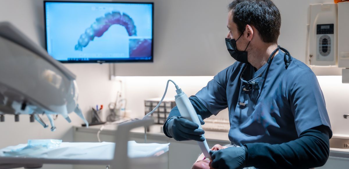
Digital dental imaging is a category of tools that capture pictures of your teeth, gums, and jaw using digital sensors instead of photographic film. Some of these tools, like digital radiographs, still use a small amount of radiation. Others, like intraoral optical scanners, use light to build a detailed model with no radiation at all.
The shared advantage is speed and clarity. Images appear on a screen within seconds, your care team can zoom in on a single tooth, and nothing has to be developed in a darkroom. That difference shapes everything from how a dental digital checkup feels to how early a problem is caught, and catching it early is the foundation of good preventative dental care.
How Digital Dental Imaging Captures Your Mouth
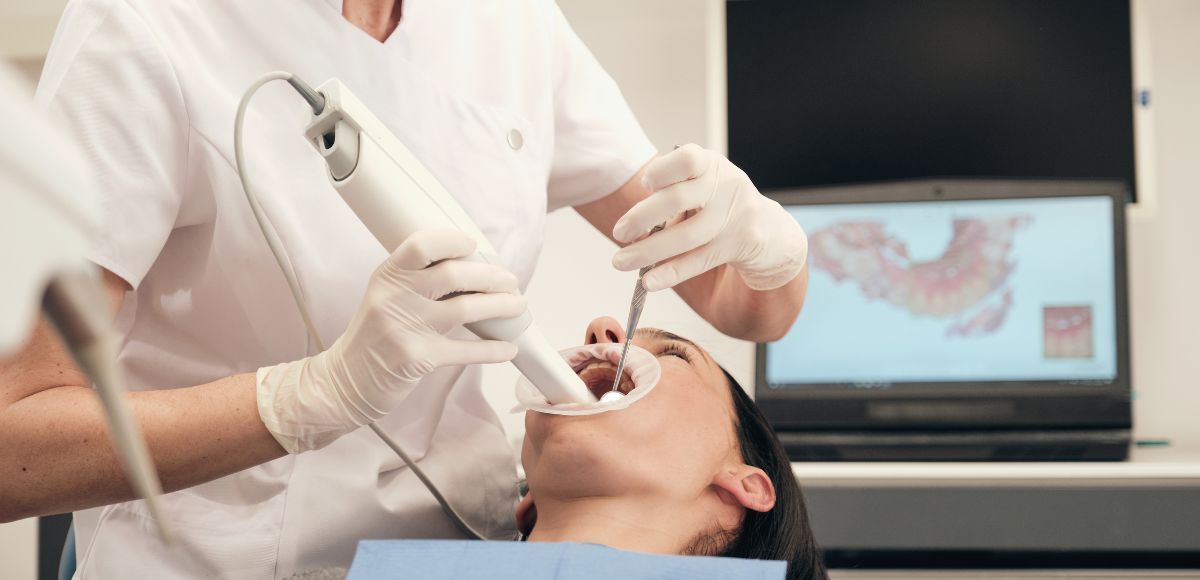
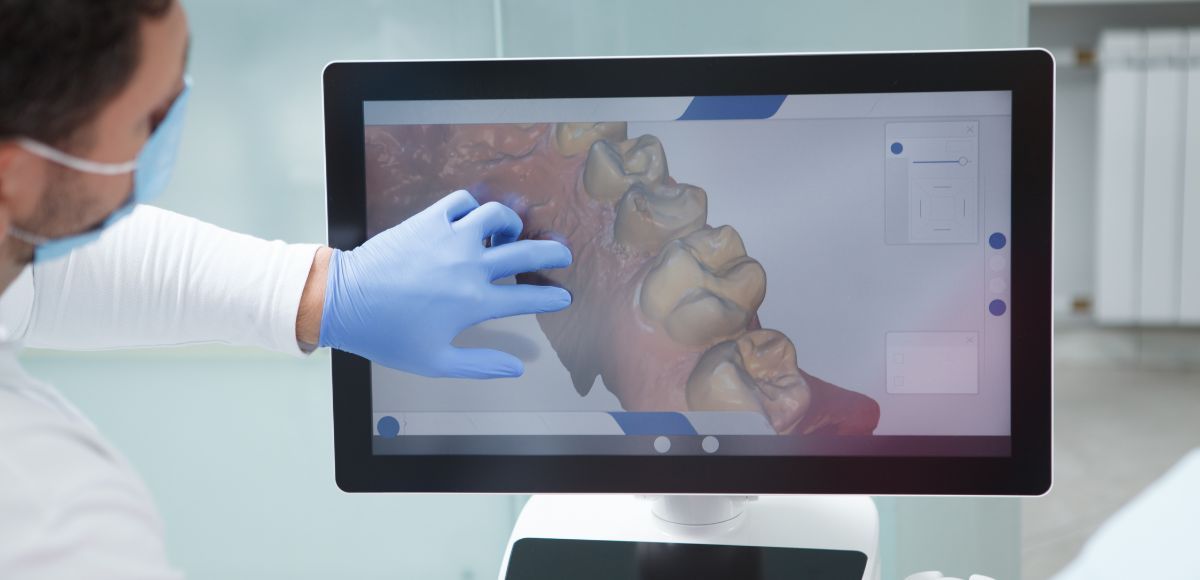
A digital sensor or a handheld wand does the work that film and bulky equipment used to do. For radiographs, a flat sensor sits where the film once went and sends the image straight to a computer. For a 3D scan, a small wand passes over your teeth and stitches a rapid sequence of frames into one accurate model. Either way, you see the result almost immediately, and so does your dentist.
How Traditional X-Rays Work and Where They Fall Short
Traditional dental X-rays rely on film that has to be chemically developed. They have served dentistry well for decades and still produce useful images. The American Dental Association's overview of dental X-rays explains how radiographs help detect decay, bone loss, and problems hidden below the gumline.
The limits show up in the details. The film takes longer to process, images cannot be enhanced or magnified on a screen, and retakes are common when the first exposure is not clear. Each retake means another round of radiation. Film also gives a flat, two-dimensional view, which can hide problems sitting at an angle the image did not capture.
Digital Imaging vs Film X-Rays: A Side-by-Side Look
When you compare digital imaging with film X-rays, the contrast is clearest across a few practical points.
None of this means film is useless. It means a 3D Dental Scan at Austin Elite Smiles gives your team more usable information from a single, comfortable appointment.
Is Digital Imaging Safer for Patients?
Safety is the question patients ask most, and it deserves a direct answer. Digital radiographs expose you to far less radiation than traditional film, often a fraction of the older dose. Optical intraoral scans, the kind used to build a 3D model, use light rather than ionizing radiation, so they add no exposure at all.
For context, the Centers for Disease Control and Prevention notes that oral health is closely tied to overall health, which is part of why catching issues early matters so much. Lower-dose imaging lets your team monitor your mouth more often without the concern that used to come with frequent film X-rays.
Accuracy: What Digital Imaging Reveals That X-Rays Can Miss
Accuracy is where digital tools change the conversation. A two-dimensional film can overlap structures, so a small cavity between two teeth or early bone loss can hide behind another surface. Digital imaging, especially 3D scanning, separates those layers and shows them clearly.
This matters for early detection. The National Institute of Dental and Craniofacial Research points out that tooth decay is largely preventable when it is found and managed early. Sharper images give your dentist a better chance to spot trouble while it is still small and simple to treat, rather than after it spreads.
Intraoral Scanning vs Radiographic Imaging: Different Tools, Different Purposes
It helps to know that not every digital tool does the same job. An intraoral scan dentist uses an optical wand to map your teeth and gums, while a full mouth scan dentist often pairs that with a low-dose radiograph for what sits underneath.
Used together, they give a complete picture that neither tool provides alone, and you can see how they fit into the wider dental services we offer.
What to Expect From Digital Imaging at Austin Elite Smiles
At our AACD-accredited practice, digital imaging is part of how we get to know your mouth before recommending anything.
New patients receive a thorough digital scan that becomes the baseline we measure future visits against. There is no film, no long wait, and no guessing about what is happening below the surface.
We use these images to explain what we see in plain language, so you understand your own health and the reasoning behind any recommendation. Many patients tell us it is the first time they have actually seen their own teeth on a screen.
If you want steady, predictable care, our Smile Wellness Plan supports consistent monitoring over time, so small changes are caught early.
Making Digital Dental Imaging Part of Your Care
Choosing between these tools is less about old versus new and more about getting the clearest, safest view of your health. Digital tools lower radiation, sharpen detail, and make it easier to catch small issues before they grow. When your dentist can see more with less exposure, you get to make calmer, better-informed decisions about your smile.
Ready to See Your Smile in Sharper Detail?
If you have been putting off a checkup because you were unsure what to expect, modern imaging makes that first visit easier than ever. Book your visit at Austin Elite Smiles and let our team show you exactly what is happening in your mouth, clearly and comfortably.
Frequently Asked Questions
1. Is digital dental imaging safer than X-rays?
Digital radiographs use far less radiation than traditional film, and optical 3D scans use none at all. For most patients, that means safer, more frequent monitoring without the radiation concerns of older film systems. Our team will always use the lowest level of imaging needed for your care
2. What is the difference between digital dental imaging and X-rays?
Traditional X-rays use film that must be developed and show a flat, two-dimensional view. Digital imaging captures the same areas with a sensor or optical wand, displays results in seconds, and allows magnification and 3D views. The practical result is faster, clearer information for both you and our team.
3. How accurate is digital dental imaging?
It is highly accurate because it separates overlapping structures and lets your dentist magnify and adjust the view. That makes early decay, bone changes, and alignment issues easier to spot than on film. Greater accuracy supports earlier, simpler treatment.
4. Do dental 3D scans use radiation?
Optical intraoral 3D scans use light to build a model, so they involve no ionizing radiation. Digital radiographs do use a small amount, but far less than traditional film. Your team chooses the right tool for each situation to keep exposure as low as possible.
5. Do I still need X-rays if I get a 3D dental scan?
An optical 3D scan and a radiograph capture different things, so one does not always replace the other. The 3D scan maps the surfaces of your teeth and your bite, while a low-dose radiograph shows roots, bone, and the areas between teeth. Our team decides which you need based on what your mouth shows at that visit, and often a combination gives the most complete view.
Key Takeaways
- Ask whether your scan is optical or radiographic. Optical 3D scans use no radiation, while digital radiographs use a small, reduced amount.
- Expect to see your images on a screen the same day, so you can understand any recommendation before agreeing to treatment.
- Treat imaging as an early-detection tool. Catching decay or bone changes early keeps care simpler.
- Ask your team to save your baseline scan so they can compare it at future visits and track subtle changes over time.
- Look for a practice that combines optical scanning with low-dose radiographs when a full picture is needed, since each tool reveals what the other cannot.